
Structure and Function of the Visual System PART 1 Biology Diagrams Aqueous humor is made by the ciliary body and flows along the posterior surface of the iris in the narrow posterior chamber. It then flows through the pupil and fills the anterior chamber. The scleral venous sinus (the old name is much more fun to say: the "canal of Schlemm") is a drainage system for the aqueous humor. Learning Objectives. By the end of this section, you should be able to. 6.1.1 Describe the region of the electromagnetic spectrum that is perceived by our visual system, and the relative energy of photons at long and short wavelengths.; 6.1.2 Describe the major parts of the eye and their role in focusing light to create a clear image.; In this section, we will meet the range of the

Both light and dark adaptation are crucial for maintaining visual function across a range of lighting conditions, demonstrating the remarkable adaptability of the human visual system. Adapted from Anatomy & Physiology by Lindsay M. Biga et al, shared under a Creative Commons Attribution-ShareAlike 4.0 International License, chapter 15.

Visual system Biology Diagrams
Humans are "Visual animals" and a relatively large proportion of human brain tissue is devoted to vision. The visual system includes the eye and retina, the optic nerves, and the visual pathways within the brain, where multiple visual centers process information about different aspects (shape and form, color, motion) of visual stimuli. + + +

The tract splits into two. The larger visual fibers head to the lateral geniculate body. The smaller fibers for the light reflex pass between the lateral and medial geniculate bodies to the midbrain. Visual fibers. The lateral geniculate body of the posterior part of the thalamus contains the third-order neurons of the visual system. Of its 6
7 Chapter 7: Sensation and Perception Biology Diagrams
Nervous System The nervous system consists of the brain, spinal cord, sensory organs, and all of the nerves that connect these organs with the rest of the body. Respiratory System The respiratory system provides oxygen to the body's cells while removing carbon dioxide, a waste product that can be lethal if allowed to accumulate. system, in combination with skilled examination, allows precise localization of neuropathological processes. Moreover, these principles guide effective diagnosis and management of neuro-ophthalmic disorders. The visual pathways perform the function of receiv-ing, relaying, and ultimately processing visual informa-tion.

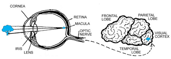
The Human Visual System. The visual system includes both the eyes and the brain. Light enters your eye where it hits the retina, which triggers light receptors to send electrical signals through your optic nerve, which travel to the back of your brain where the first stages of visual perception take place. visual system" /> Source:https
